Publication 2022 – VetAgro Sup

Use of a silicone skin extension medical device
for equine wound management

Lepage Olivier M, Paindaveine Charlotte, Verkaede Marlies
Groupe de Recherche en Médecine et Rééducation des Equidés de Sport (GREMERES), Centre for Equine Health, Veterinary School of Lyon, VetAgro Sup, University of Lyon, France
Introduction
- In horses, second-line repair of a traumatic limb wound often leads to complications, including the development of exuberant granulation tissue that can produce a chronic wound that cannot heal.[1]
- When the integrity of the horse’s skin is broken, the interaction between cellular and molecular events, which are the source of the dynamic healing process, is a little different from other species.[2]
- Beyond the simple anatomical location of an injury, the horse’s size, weight, character and physical activity are major constraints that must be taken into account when prescribing treatment.
- A multitude of treatments such as dressings specific to the different phases of healing or the application of creams containing mesenchymal stem cells (or their secretome) are used [3].
- The use of a tissue extension under uni-axial traction has been described in humans to reduce tension at the edges of a wound [4].
- Reports of a skin extension medical device made from silicone are given here for a series of clinical cases in horses.
Materiel & Method
- Prospective and descriptive study of horses presented for wound management on which a MID-VET™ silicone device (Medical Innovation Development S.A.S., Dardilly, France) was used between September 2020 and July 2021.
- The silicone skin extension device is composed of a 100 cm wire, a swaged curved reversed cutting needle, 8 blocks and 4 tension adjustment blockers.
- Type of treatment : The device was placed during surgery under general anaesthesia. A same clinician in a same hospital setting (Clinéquine, VetAgro Sup) followed all horses.
- Technique : The silicone wire was moistened before use and not tightened until the swaged needle has been cut. Sutures were placed so that the blocks were not in contact with the wound. The tension was readjusted as needed using the reversible adjustment blockers.
Results
- The device has been implanted six times (four adult horses aged between 5 and 12 years and two fillies aged 10 days and 2 months)
- Indications: acute trauma with
1) Loss of skin substance requiring partial or total secondary intention wound healing (n=4),
(2) Significant wound tension (n=2). - Complications: silicone blocks became embedded in the skin, with no consequences on wound healing (n=4); and with necrosis and tear formation of the wound margin (n=2).
- Duration of use before removal: 7 to 11 days
- Impact on wound progression: approximately one week after application, the device allows granulation tissue from the wound bed to adhere to its margins.
Discussion and clinical significance
- The MID-VET™ device is easy to use in young and adult horses.
- Avoid putting too much tension on the wire during application and do not exceed double the length of the elastic silicone wire to avoid skin necrosis.
- The silicone device facilitates second intention wound healing and helps reduce tension when first intention wound healing is expected in a complicated anatomical area.
- For each case, the potential contribution of the device to the healing process must be assessed in terms of the additional cost associated with the device.
Wounds with significant tension (n=2)
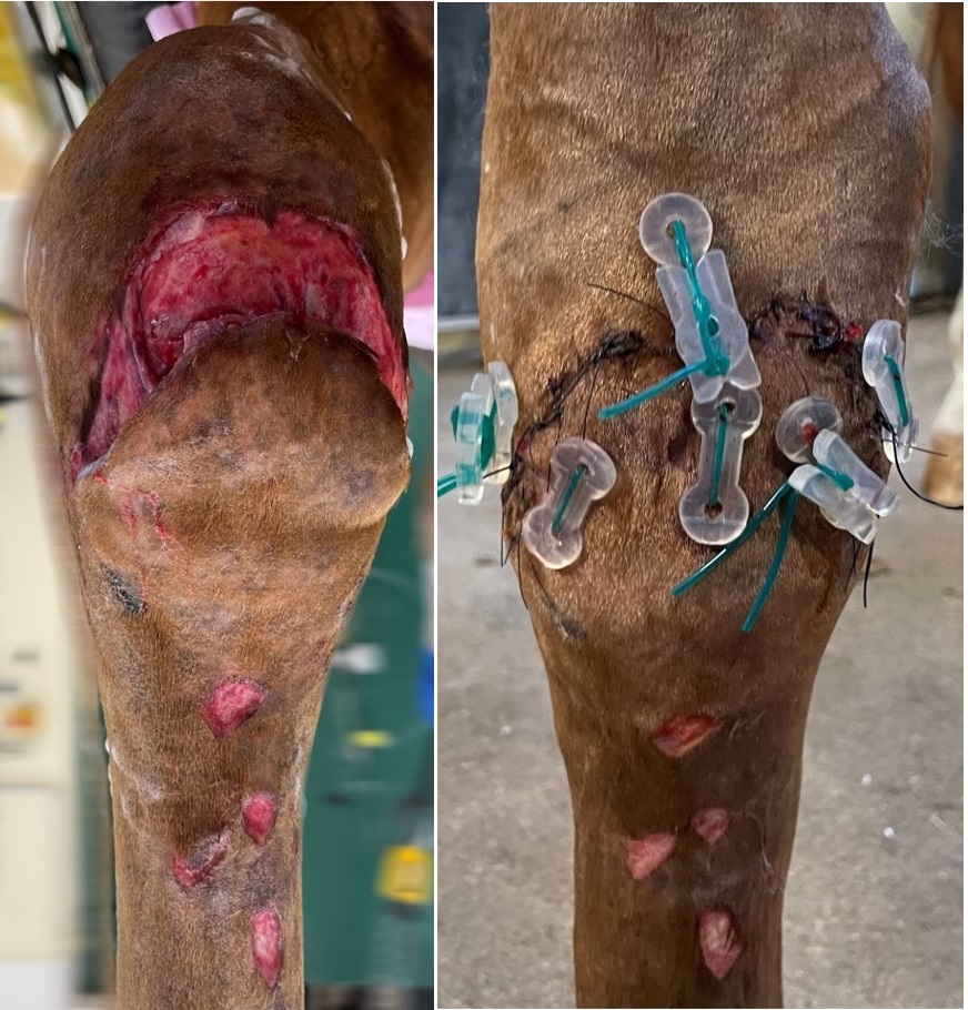
Figure 1: Twelve-year-old SF gelding who escaped from the owner and was found after two days with multiple injuries. On the right carpus after debridement and lavage of the wound and the synovial sheaths, reconstruction was carried out to obtain first-line healing with the help of a MID-VET™ device.
The device was removed on the tenth day.
First intention wound healing took place without any problems despite the significant tension on the dorsal surface of this joint.
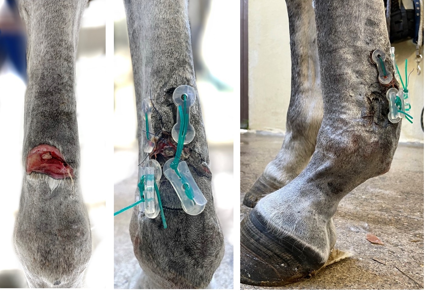
Figure 2: Six-year-old Thoroughbred mare escaped from the meadow after pulling out stakes and wires. Admitted with multiple wounds including one in the palmar region of the left front limb. After debridement and lavage of the wound, a MID-VET™ device was applied to reduce tension at the edges of the wound, which were connected with simple interrupted suture pattern.
The silicone device was removed on day 11 and the rest of the sutures on day 13. Primary wound healing was uneventful.
Second intention wound healing (n=4)
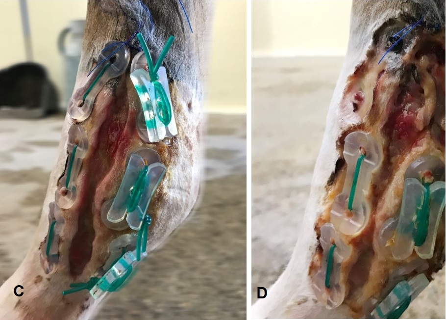
Figure 3: Two-month-old SF filly with a wound on the right front fetlock which occurred in a field approximately 18 hours before surgery. (A) A significant retraction of the skin around the joint was noted; (B) three horizontal mattress sutures were made using the elastic silicone suture material passed through two silicone blocks positioned on each side of the wound without touching the wound. The blocks have two holes for the suture material to pass through and a tension adjustment blocker on one side. Six and nine days post-surgery, good reduction of the wound (C) and skin necrosis under the blocks (D) were respectively observed.
The device was removed on the day nine and second intention wound healing continued uneventful.
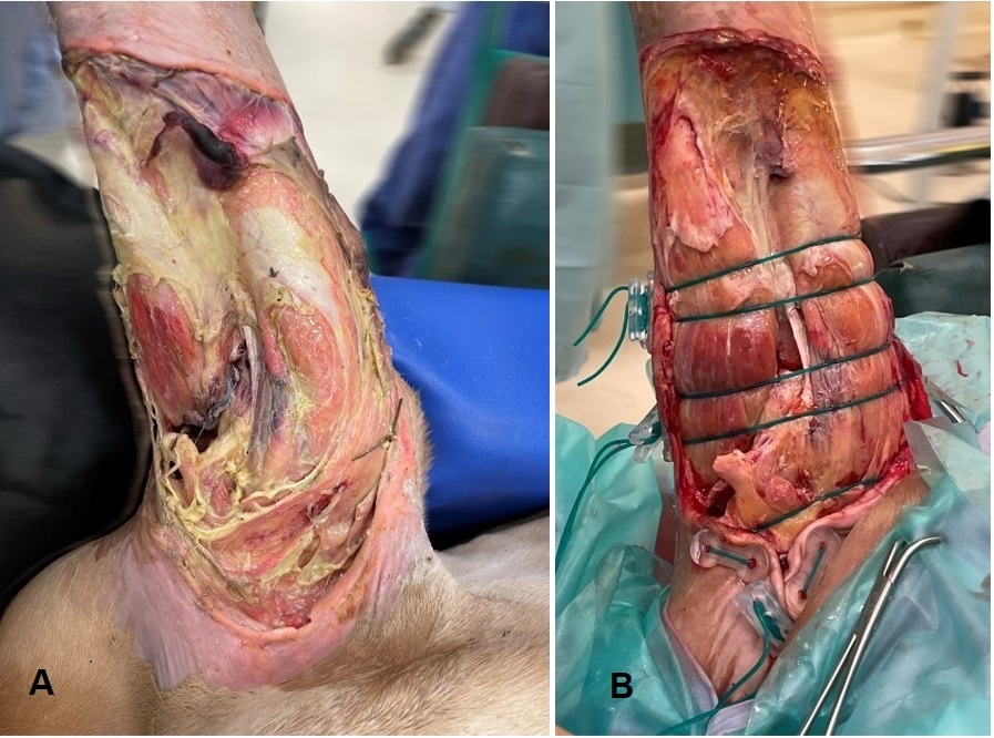
Figure 4: SF filly less than 12-hours-old found in a barbed wire fence and admitted with multiple wounds, one of which involved the left front limb from the elbow to the carpus.
(A) total absence of soft skin with multiple deep vascular lesions and retraction of healthy skin on the lateral side of the limb; (B) a MID-VET™ device was applied with eight blocks.
The device was removed on day eight; second intention wound healing continued its normal process but the filly died seven days later from metabolic disorders.
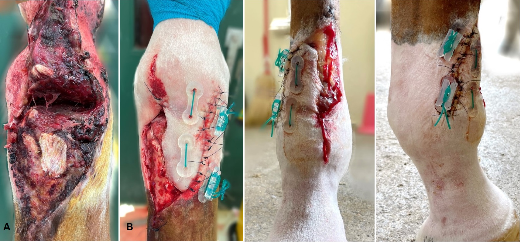
Figure 5 : Seven-year-old SF gelding injured in a field about 18 hours before surgical treatment and admitted with (A) a wound on the left hind fetlock with complete section of the extensor digitorum communis and an area of skin necrosis. (B) After debridement and lavage of the wound, a MID-VETTM device was placed medially to reduce tension on the skin flap intended to cover this high motion joint.
The silicone sutures were removed after seven days, after which perfect adhesion of the skin flap combined with a reduction in the second intention wound healing surface was observed.
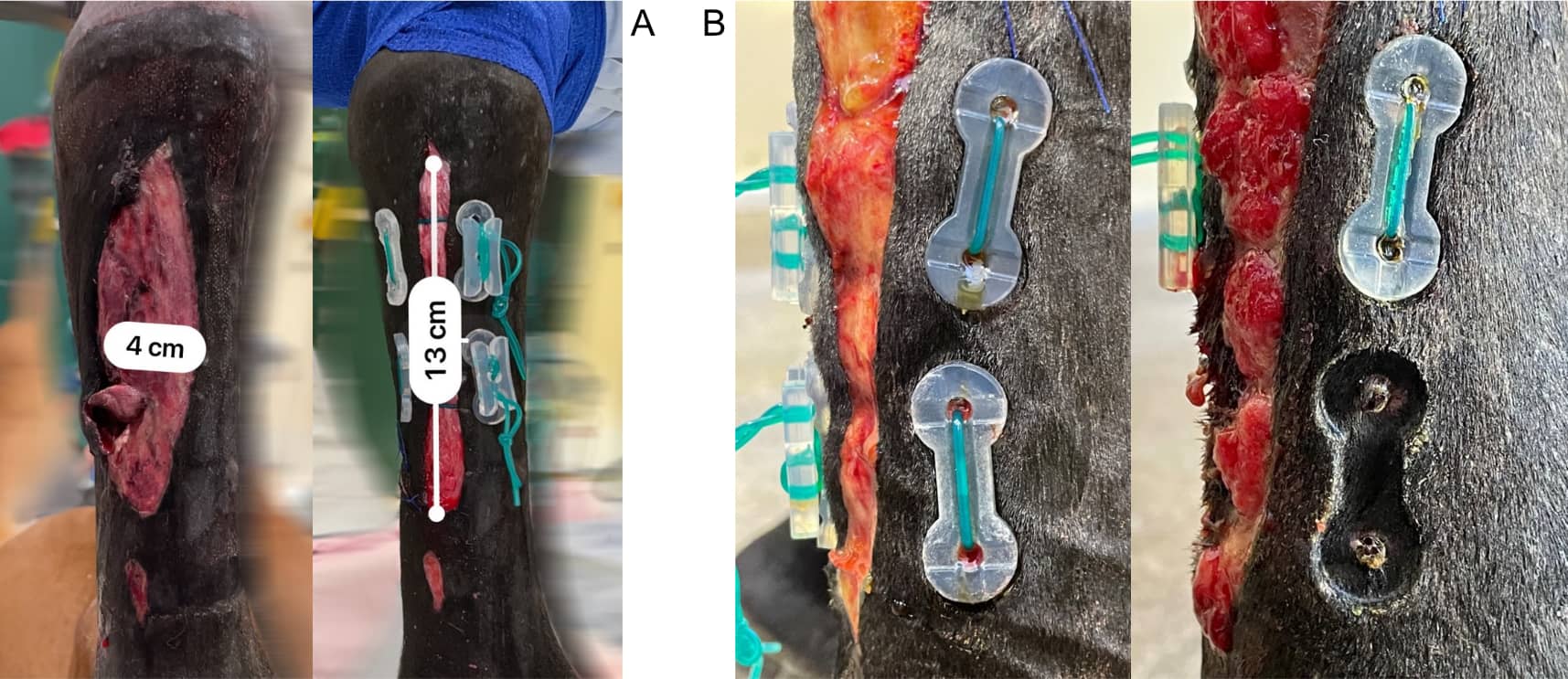
Figure 6 : Eight-year-old Arabian mare admitted for lameness and a major wound in regard of the left front metacarpal region. (A) A silicone skin extension medical device was applied to cover the area of the tendons as much as possible and to speed up second intention wound healing; (B) The device was removed after 11 days.
The skin under the blocks had been crushed, but there was no necrosis and no consequences have been observed for the rest of the healing process .
Conflicts of interest : The authors declare no conflict of interest but that these data are
taken from clinical cases for which Medical Innovation Development SAS sponsored the
silicone material.
1. Stashak T, Theoret C. Equine Wound Management, Wiley- Blackwell, Ames, LA, USA, 2008.
2. Wilmink JM, van Weeren PR. Second-intention repair in the horse and pony and management of exuberant granulation tissue, Veterinary Clinics of North America: Equine Practice, vol. 21, no. 1, pp. 15–32, 2005.
3. Di Francesco P, Cajon P, Desterke C, Perron Lepage MF, Lataillade J-J, Kadri T, Lepage OM. Effect of Allogeneic Oral Mucosa Mesenchymal Stromal Cells on Equine Wound Repair. Vet Med Intern , doi.org/10.1155/2021/50249052021
4. Simon E. Etude des possibilités d’extension tissulaire sous l’effet d’une traction uni-axiale et applications cliniques. Thèse Université Nancy 1-Henri Poincaré UFR Médecine, 2005.
EWMA & VWHA International Conference, Paris 2022